Friday Factoid: CT Scan v MRI
What's the difference?
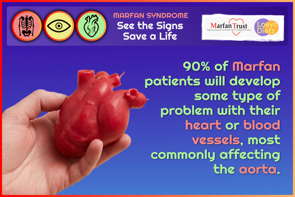
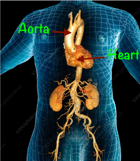
-Both of these imaging modalities are frequently used in individuals with Marfan syndrome.
- Your doctors will carefully assess the risks and benefits of both types of imaging to choose the type of scan that is best suited to you
-These scans are often done alongside other tests like echocardiograms which may be carried out more regularly
-It's helpful for doctors to compare 'like with like' so they will often try and repeat the same type of test for comparison purposes
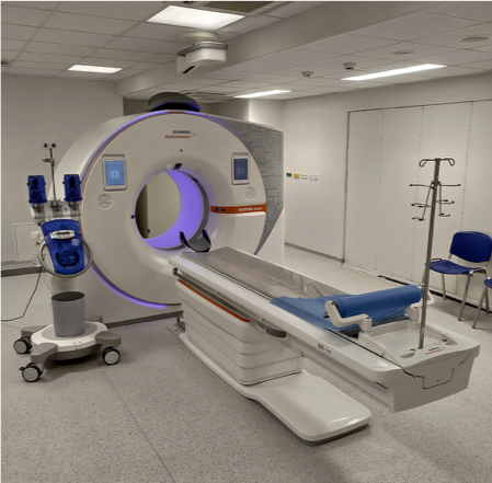
Fast (usually under 15 minutes)
Contrast may be used to improve pictures
Better for acute/emergency situations like aortic dissection
Widely available nationwide
Uses magnets and radio waves to obtain 3D images of your body structures
Slower (usually 30-60 minutes)
Contrast may be used to improve pictures
Better for long term aortic monitoring and repeated imaging (due to no use of radiation)
May need to travel to a specialist centre for detailed cardiac/aortic imaging
Can be claustrophobic
Need to make staff aware of any metal in the body e.g. pacemakers