Connective Cousins
The stories behind four connective tissue disorders

Dr Antoine Marfan
In 1896, Dr Antoine Marfan, a French paediatrician working in the Faculty of Medicine in the University of Paris, presented a young girl called Gabrielle P to his colleagues. Gabrielle's physique was characterised by long, slender limbs and joint contractures. He called the condition dolichostenomelia (which is Greek for slender limbs).
Six years later, in 1902, Gabrielle P was examined using the new technology of X-rays by Drs Henri Mery and Leon Babonneix. She was noted to have abnormalities of her thoracic spine and her diagnosis was changed to 'hyperchondroplasia', the opposite of the condition ‘achondroplasia’.
That same year, Dr Emile Achard reported a patient with similar physical features and used the term ‘arachnodactylie’. Dr Achard described his patient as hypermobile and noted a positive family history of similar features.
Several people with similar features and, importantly, complications affecting the eye or aorta, were described in the early part of the 20th century, but the term ‘Marfan syndrome’ was only proffered for the first time (in the journal ‘Le Nourisson’ – ‘the infant’) in 1929 by Dr Carrau (who used the term ‘maladie de Marfan’).
Ironically, it was probably Dr Achard who described the first case of what we now call Marfan syndrome and Gabrielle P may not have had Marfan syndrome at all, but a related condition now known as Beal’s syndrome (or congenital contractural arachnodactyly).
In the 1950s, Dr Victor McKusick summarised the collective experience of many different clinicians and wrote what is probably the first modern account of Marfan syndrome in his momentous publication ‘Heritable Disorders of Connective Tissue’. He focused on the link between lens dislocation and aortic aneurysm.
Since the 1950s, diagnostic criteria for Marfan syndrome have been refined and, in 1991, our medical director, Dr Child pioneered the international consortium that discovered the causative gene for Marfan syndrome - FBN1. With more known about the genetics of the condition, medical treatment has correspondingly improved. As the late physician, Dr Reed Pyeritz, remarked: "thirty years of research into Marfan syndrome equals thirty years of additional life expectancy".
Dr Marfan went on to become a specialist in children’s infectious disease and hygiene.
Gabrielle P died in adolescence, possibly from tuberculosis.
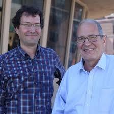
Dr Bart Loeys is a prominent geneticist based in Belgium who specialises in rare genetic connective tissue disorders and cardiovascular genetics. Meanwhile, Dr Hal Dietz is a renowned paediatric cardiologist and professor at John Hopkins Medicine in the United States. Together with Dr Loeys he described Loeys-Dietz in 2005. Prior to its identification, many patients with this condition were thought to have Marfan syndrome.
The doctors noticed a group of patients with severe arterial aneurysms (widened blood vessels) who did not quite fit the standard diagnosis for Marfan syndrome They discovered that the condition was caused by mutations in the genes encoding transforming growth factor-beta receptors (TGFBR1 and TGFBR2), which disrupt the body's normal connective tissue signaling pathway. For more on this condition click here.
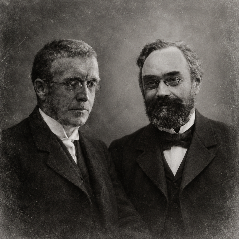
The story of Ehlers-Danlos syndrome stretches back to Hippocrates, who described people with unusually loose joints as early as 400 BC. Centuries later, Danish dermatologist Edvard Ehlers (1863 – 1937) and French physician Henri-Alexandre Danlos (1844 – 1912) independently described the distinctive features of the condition that now bears their names at the turn of the twentieth century. Today, Ehlers-Danlos syndromes encompass a group of genetic connective tissue disorders, characterised by features such as joint hypermobility, chronic pain, stretchy or fragile skin and abnormal scarring, aortic dissection with symptoms and severity varying considerably between types.
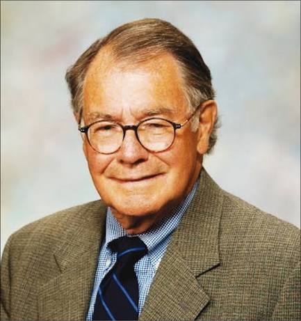
In 1960, paediatrician Gunnar Stickler examined a 12-year-old boy at a children’s clinic in Minnesota who had unusually enlarged joints and was extremely short-sighted. His mother was blind, and when Stickler looked more closely, he discovered other members of the family with similar features. Intrigued by this pattern, he began studying the family with his colleagues, gradually joining the dots between the problems affecting their eyes and joints. Their findings were published in 1965, describing what Stickler tentatively called ‘hereditary progressive arthro-ophthalmopathy’ - the condition we now know as Stickler syndrome. For more information click here and here.